 EN
EN

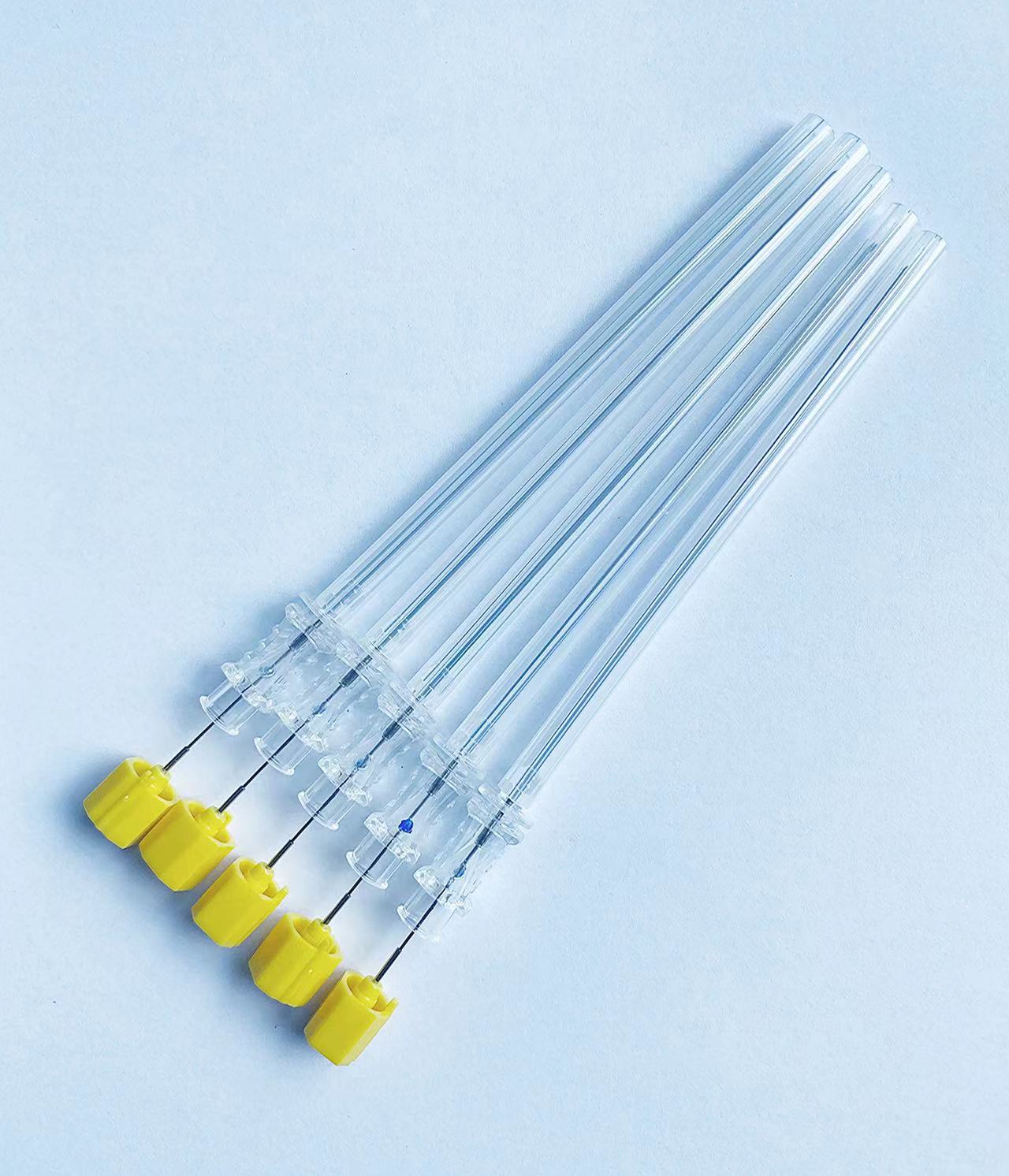
A spinal needle serves as one of the most critical medical instruments in modern healthcare, specifically designed for accessing the subarachnoid space within the spinal column. This specialized medical device enables healthcare professionals to perform essential procedures such as lumbar punctures, spinal anesthesia, and cerebrospinal fluid collection with precision and safety. Understanding the comprehensive applications and proper usage of a spinal needle is fundamental for medical practitioners working in anesthesiology, neurology, emergency medicine, and surgical specialties.

Primary Medical Applications of Spinal Needles
Diagnostic Lumbar Puncture Procedures
Medical professionals utilize a spinal needle primarily for diagnostic lumbar punctures, a procedure that allows direct access to cerebrospinal fluid for laboratory analysis. This diagnostic technique proves invaluable in identifying various neurological conditions including meningitis, encephalitis, subarachnoid hemorrhage, and multiple sclerosis. The spinal needle must penetrate through skin, subcutaneous tissue, muscle, and ligaments to reach the subarachnoid space between the third and fourth lumbar vertebrae.
During diagnostic procedures, healthcare providers carefully insert the spinal needle while monitoring patient responses and cerebrospinal fluid flow characteristics. The collected fluid undergoes comprehensive laboratory testing including cell count analysis, protein measurements, glucose levels, and microbiological cultures. Proper spinal needle technique ensures accurate sample collection while minimizing patient discomfort and potential complications such as post-puncture headaches or infection risks.
Therapeutic Spinal Interventions
Beyond diagnostic applications, a spinal needle facilitates numerous therapeutic interventions including intrathecal medication administration and cerebrospinal fluid pressure reduction. Physicians employ spinal needles for delivering chemotherapy drugs directly into the central nervous system, administering antibiotics for severe infections, and providing pain management medications for chronic conditions. These therapeutic applications require precise needle placement and careful monitoring throughout the procedure.
Therapeutic spinal punctures often involve removing excess cerebrospinal fluid to relieve increased intracranial pressure associated with conditions like idiopathic intracranial hypertension or normal pressure hydrocephalus. The spinal needle allows controlled fluid drainage while maintaining sterile technique and patient safety protocols. Medical teams must carefully balance fluid removal rates with patient hemodynamic stability during these critical interventions.
Spinal Anesthesia and Regional Block Applications
Surgical Anesthesia Administration
Anesthesiologists extensively use spinal needles for administering spinal anesthesia during various surgical procedures, particularly those involving the lower abdomen, pelvis, and lower extremities. The spinal needle enables precise delivery of local anesthetic agents into the subarachnoid space, creating temporary numbness and paralysis below the injection site. This technique provides excellent surgical anesthesia while allowing patients to remain conscious during procedures.
Spinal anesthesia offers numerous advantages over general anesthesia including reduced respiratory complications, decreased blood loss, improved postoperative pain control, and faster recovery times. The spinal needle must be positioned accurately to ensure proper anesthetic distribution while avoiding potential complications such as nerve damage or epidural hematoma formation. Anesthesiologists carefully select needle gauge and tip design based on patient anatomy and procedural requirements.
Obstetric and Gynecologic Procedures
Obstetric anesthesia represents one of the most common applications for spinal needle usage, particularly for cesarean section deliveries and labor pain management. The spinal needle allows rapid onset of surgical anesthesia while maintaining maternal consciousness and facilitating immediate bonding with newborns. Proper spinal needle technique in obstetric patients requires special considerations for anatomical changes during pregnancy and potential effects on fetal circulation.
Gynecologic procedures including hysterectomies, ovarian surgeries, and pelvic reconstructive operations frequently utilize spinal anesthesia delivered through carefully positioned spinal needles. The technique provides excellent muscle relaxation and surgical conditions while minimizing systemic drug exposure and associated side effects. Anesthesiologists must consider patient positioning, surgical duration, and postoperative pain management when planning spinal needle interventions for gynecologic cases.
Technical Specifications and Design Features
Needle Gauge and Length Considerations
Spinal needle selection requires careful consideration of gauge size, length, and tip configuration based on specific procedural requirements and patient characteristics. Common spinal needle gauges range from 18 to 27 gauge, with smaller numbers indicating larger diameter needles that allow faster cerebrospinal fluid flow but potentially increase patient discomfort and complication risks. The spinal needle length varies typically between 3.5 and 6 inches, accommodating different patient body habitus and anatomical variations.
Healthcare providers must balance multiple factors when selecting appropriate spinal needle specifications including procedure type, patient age, body mass index, and anticipated technical difficulties. Larger gauge spinal needles facilitate rapid cerebrospinal fluid drainage but may cause more tissue trauma and increase headache incidence. Conversely, smaller gauge needles reduce patient discomfort but require longer procedure times and may become occluded by tissue debris or blood products.
Tip Design and Bevel Configuration
Modern spinal needle designs incorporate various tip configurations including pencil-point, cutting bevel, and blunt-tip options, each offering distinct advantages for specific applications. Pencil-point spinal needles feature rounded tips that separate dural fibers rather than cutting them, potentially reducing cerebrospinal fluid leakage and post-procedural headache incidence. These needles require slightly more insertion force but provide excellent tactile feedback during advancement through tissue layers.
Cutting bevel spinal needles utilize sharp, angled tips that slice through tissues with minimal resistance, facilitating easier insertion in challenging cases or patients with calcified ligaments. However, these needles may cause more tissue trauma and potentially increase complication rates compared to pencil-point designs. The spinal needle bevel orientation affects cerebrospinal fluid flow patterns and must be carefully positioned to optimize drainage while minimizing dural tear size.
Safety Protocols and Best Practices
Sterile Technique and Infection Prevention
Proper spinal needle procedures demand strict adherence to sterile technique protocols to prevent potentially catastrophic complications such as meningitis or epidural abscess formation. Healthcare providers must perform thorough hand hygiene, don sterile gloves and gowns, and utilize sterile draping techniques before handling any spinal needle components. The insertion site requires comprehensive skin preparation using appropriate antiseptic solutions and adequate drying time for maximum antimicrobial effectiveness.
Single-use, disposable spinal needles eliminate risks associated with inadequate sterilization or equipment reprocessing errors. Medical facilities must maintain proper storage conditions for spinal needle inventory, ensuring package integrity and expiration date compliance. Healthcare teams should establish clear protocols for spinal needle handling, including proper disposal methods for contaminated equipment and documentation of all procedural details for quality assurance purposes.
Patient Positioning and Anatomical Landmarks
Successful spinal needle placement requires optimal patient positioning and accurate identification of anatomical landmarks to ensure safe, effective procedures. The lateral decubitus position with knees drawn toward the chest maximizes intervertebral space opening, while the sitting position may facilitate landmark identification in obese patients or those with challenging anatomy. Healthcare providers must carefully palpate bony landmarks including the iliac crests and spinous processes to determine appropriate spinal needle insertion sites.
Proper spinal needle insertion technique involves advancing the needle through progressive tissue layers while maintaining appropriate angle and depth control. Practitioners should recognize characteristic changes in tissue resistance and monitor for cerebrospinal fluid return indicating successful subarachnoid space entry. Patient comfort measures including local anesthetic infiltration and clear communication throughout the procedure help minimize anxiety and movement that could compromise spinal needle placement accuracy.
Complications and Risk Management
Immediate Procedural Complications
Spinal needle procedures carry inherent risks that healthcare providers must recognize and manage appropriately to ensure optimal patient outcomes. Immediate complications may include vasovagal reactions, bleeding at the insertion site, nerve root irritation, or inadvertent dural puncture during epidural procedures. Proper spinal needle technique and careful patient monitoring help identify early warning signs of complications before they progress to more serious conditions.
Healthcare teams must maintain emergency equipment and medications readily available during all spinal needle procedures, including resuscitation supplies, vasopressor agents, and airway management tools. Prompt recognition and treatment of complications such as total spinal anesthesia or cardiovascular collapse require coordinated team responses and clear communication protocols. Regular training and simulation exercises help maintain staff proficiency in managing spinal needle-related emergencies.
Long-term Complications and Follow-up Care
Post-procedural complications following spinal needle use may include persistent cerebrospinal fluid leakage, chronic headaches, or rarely, neurological deficits requiring extended medical management. Healthcare providers should educate patients about potential warning signs and appropriate follow-up care after spinal needle procedures. Clear discharge instructions must include activity restrictions, symptom monitoring guidelines, and emergency contact information for urgent concerns.
Long-term complication prevention involves careful spinal needle technique, appropriate patient selection, and thorough informed consent processes that explain risks and benefits of proposed interventions. Medical documentation should include detailed procedure notes, patient responses, and any complications encountered during spinal needle placement. Quality improvement initiatives help identify patterns in complication rates and guide modifications to procedural protocols or equipment selection.
FAQ
What conditions require the use of a spinal needle for diagnosis
A spinal needle is essential for diagnosing various neurological conditions through cerebrospinal fluid analysis. These include suspected meningitis, encephalitis, subarachnoid hemorrhage, multiple sclerosis, Guillain-Barre syndrome, and certain cancers affecting the central nervous system. The spinal needle allows healthcare providers to collect cerebrospinal fluid samples for laboratory testing, measuring cell counts, protein levels, glucose concentrations, and identifying infectious organisms or malignant cells.
How painful is a procedure involving a spinal needle insertion
Most patients experience only mild to moderate discomfort during spinal needle procedures when proper local anesthesia is administered beforehand. The initial skin injection of local anesthetic causes brief stinging, followed by numbness that significantly reduces pain during actual spinal needle insertion. Patients typically describe pressure sensations rather than sharp pain, and the entire procedure usually takes 15-30 minutes. Healthcare providers use various comfort measures including positioning aids and continuous communication to minimize patient anxiety and discomfort.
What is the difference between spinal and epidural needle procedures
A spinal needle penetrates completely through the dura mater into the subarachnoid space containing cerebrospinal fluid, while epidural needles stop in the epidural space outside the dura. Spinal needle procedures typically use smaller gauge needles and provide faster onset of anesthesia with smaller medication volumes, but effects cannot be easily adjusted once administered. Epidural procedures allow continuous medication administration through catheter placement but require larger needles and more complex technique for proper positioning.
How long do the effects of spinal anesthesia administered through a spinal needle last
The duration of spinal anesthesia depends on the specific medications injected through the spinal needle, typically lasting 2-6 hours for surgical procedures. Local anesthetics like bupivacaine provide longer-lasting effects compared to lidocaine, while additives such as opioids or epinephrine can extend anesthesia duration. Motor function usually returns before sensory function, with complete recovery expected within 6-24 hours depending on the specific spinal needle technique and medications used. Healthcare providers carefully time spinal needle procedures to match anticipated surgical duration while ensuring patient safety throughout the recovery period.