 EN
EN

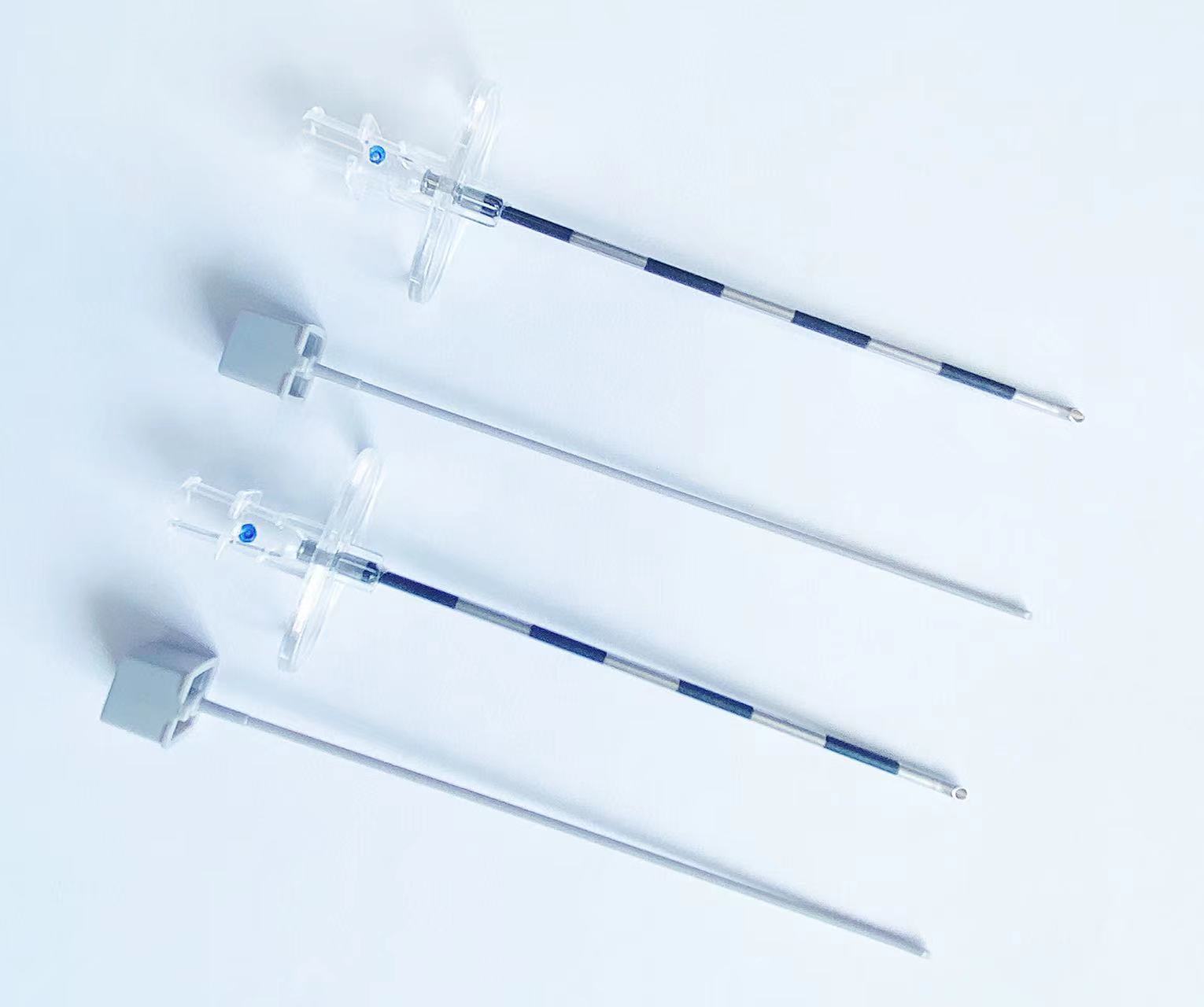
Selecting the optimal spinal needle for fluoroscopic-guided procedures represents a critical decision that directly impacts injection precision, patient safety, and procedural success rates. The visibility characteristics of different spinal needle designs under fluoroscopic imaging can significantly influence the accuracy of targeted spinal injections, particularly in complex anatomical regions where precise needle placement is paramount for therapeutic efficacy and complication avoidance.

Fluoroscopic visibility in spinal needle selection depends on multiple technical factors including needle gauge, tip design, material composition, and surface treatment characteristics. Understanding these variables enables clinicians to make informed decisions about which spinal needle configuration provides superior radiographic contrast and positioning accuracy for specific injection procedures, ultimately enhancing patient outcomes and procedural confidence in challenging anatomical scenarios.
Needle Design Features That Enhance Fluoroscopic Visibility
Material Composition and Radiopacity Characteristics
The fundamental radiopacity of a spinal needle depends primarily on its material composition and metallic density characteristics. Stainless steel spinal needles typically provide excellent fluoroscopic contrast due to their high atomic number and density properties, creating clear radiographic shadows that facilitate precise positioning under real-time imaging guidance. The material thickness and alloy composition directly influence the degree of X-ray absorption, with higher density materials producing more pronounced visibility on fluoroscopic displays.
Advanced spinal needle manufacturing incorporates specific alloy compositions designed to optimize radiographic contrast without compromising structural integrity or biocompatibility requirements. These specialized materials ensure that the spinal needle maintains consistent visibility characteristics throughout the injection procedure, even when navigating through dense bone structures or complex anatomical planes where standard needles might become difficult to visualize effectively.
Tip Configuration and Geometric Design Impact
The geometric design of the spinal needle tip significantly affects its fluoroscopic presentation and positioning accuracy during targeted injection procedures. Pencil-point spinal needle designs often provide superior tip visualization compared to cutting needles due to their rounded configuration, which creates a more defined radiographic silhouette that remains clearly visible even when positioned at challenging angles within the spinal anatomy.
Beveled spinal needle tips present different visibility characteristics depending on their orientation relative to the fluoroscopic beam, with the cutting edge creating variable contrast patterns that experienced clinicians can use to assess needle rotation and precise positioning. The tip geometry also influences how the spinal needle interacts with surrounding tissues, affecting both procedural accuracy and patient comfort during complex injection procedures.
Gauge Selection Considerations for Optimal Visibility
Large Gauge Needles and Enhanced Contrast
Larger gauge spinal needles generally provide superior fluoroscopic visibility due to their increased cross-sectional area and greater material volume, which results in enhanced X-ray absorption and more pronounced radiographic contrast. A 18-gauge spinal needle typically offers excellent visibility characteristics that facilitate precise positioning even in challenging anatomical locations, making it suitable for procedures requiring maximum positioning accuracy and real-time visualization feedback.
The enhanced visibility of large gauge spinal needles comes with trade-offs in terms of tissue trauma and patient comfort, requiring clinicians to balance optimal imaging characteristics against procedural invasiveness. However, for complex procedures where precise needle placement is critical for therapeutic success and complication avoidance, the superior fluoroscopic visibility of larger gauge needles often justifies their use despite the increased tissue disruption.
Fine Gauge Needles and Visibility Challenges
Fine gauge spinal needles, while offering advantages in terms of patient comfort and tissue preservation, present significant challenges for fluoroscopic visualization due to their reduced material volume and decreased X-ray absorption characteristics. A 25-gauge spinal needle requires optimal fluoroscopic technique and potentially enhanced imaging parameters to maintain adequate visibility throughout the injection procedure, particularly when navigating through dense anatomical structures.
Compensating for reduced visibility in fine gauge spinal needles often requires enhanced fluoroscopic techniques including increased beam intensity, optimized collimation, and careful attention to patient positioning to minimize interference from overlying structures. Advanced imaging systems with digital enhancement capabilities can significantly improve the visualization of fine gauge spinal needles, making them more practical for precision injection procedures where minimal tissue trauma is prioritized.
Surface Treatment Technologies for Enhanced Visualization
Specialized Coatings and Marking Systems
Modern spinal needle manufacturing incorporates specialized surface treatments and marking systems designed to enhance fluoroscopic visibility without compromising biocompatibility or procedural safety. Radiopaque markers positioned at specific intervals along the needle shaft provide depth reference points that assist clinicians in determining precise insertion depth and positioning accuracy during complex injection procedures.
Advanced coating technologies can enhance the natural radiopacity of spinal needle materials while maintaining smooth surface characteristics that facilitate easy tissue penetration and reduced procedural friction. These specialized treatments often incorporate biocompatible materials with high atomic numbers that significantly improve fluoroscopic contrast without adding substantial weight or altering the mechanical properties of the spinal needle.
Tip Enhancement Technologies
Specialized tip enhancement technologies focus on improving the visibility of the most critical portion of the spinal needle during precise positioning procedures. Some manufacturers incorporate radiopaque tip materials or specialized geometric configurations that create enhanced contrast at the needle point, where accurate positioning is most crucial for procedural success and patient safety.
These tip enhancement approaches must balance improved visibility with maintained sharpness and penetration characteristics, ensuring that enhanced fluoroscopic properties do not compromise the fundamental performance requirements of the spinal needle during tissue penetration and precise positioning procedures. Advanced manufacturing techniques enable the integration of visibility enhancement features without sacrificing the essential functional characteristics required for successful spinal injection procedures.
Procedural Technique Optimization for Maximum Visibility
Fluoroscopic Parameter Adjustment
Optimizing fluoroscopic parameters represents a critical component in maximizing spinal needle visibility regardless of the specific needle design characteristics. Proper beam collimation, appropriate kilovoltage settings, and optimal patient positioning can significantly enhance the visibility of any spinal needle configuration, making even fine gauge needles clearly visible during complex injection procedures.
Advanced fluoroscopic systems offer digital enhancement capabilities that can amplify the contrast characteristics of spinal needles in real-time, providing improved visualization without requiring changes to needle selection or procedural technique. These digital enhancement features are particularly valuable when working with fine gauge spinal needles or when operating in anatomical regions with challenging visualization characteristics.
Anatomical Positioning and Approach Angle Optimization
The approach angle and patient positioning significantly influence spinal needle visibility under fluoroscopic guidance, with optimal positioning enhancing visibility regardless of the specific needle characteristics. Proper patient positioning can minimize interference from overlying bone structures and optimize the radiographic presentation of the spinal needle throughout the injection procedure.
Understanding the relationship between fluoroscopic beam angle, patient anatomy, and needle positioning enables clinicians to optimize visibility for any spinal needle configuration. Strategic approach planning can compensate for inherent visibility limitations in specific needle designs, ensuring adequate visualization throughout even complex injection procedures in challenging anatomical locations.
Clinical Decision Framework for Needle Selection
Procedure-Specific Visibility Requirements
Different spinal injection procedures have varying visibility requirements that influence optimal spinal needle selection for fluoroscopic guidance. Epidural injections in the lumbar region may benefit from larger gauge needles with enhanced visibility characteristics, while precise facet joint injections might require fine gauge needles despite their reduced fluoroscopic presence, necessitating compensatory imaging techniques.
Complex procedures such as selective nerve root blocks or discography require exceptional positioning accuracy that often favors spinal needles with superior visibility characteristics, even when this means accepting increased tissue trauma or procedural invasiveness. The clinical judgment regarding visibility versus other procedural factors requires careful consideration of patient-specific factors and procedural complexity requirements.
Patient-Specific Factors Affecting Needle Choice
Patient body habitus, bone density, and anatomical variations significantly influence the optimal spinal needle selection for fluoroscopic-guided procedures. Obese patients or those with dense bone structures may require larger gauge spinal needles with enhanced visibility characteristics to maintain adequate positioning accuracy throughout the injection procedure.
Elderly patients with osteoporotic changes may benefit from fine gauge spinal needles despite their reduced visibility, as the decreased bone density can actually improve overall fluoroscopic contrast and make smaller needles more visible than they would be in younger patients with denser bone structures. These patient-specific considerations must be balanced against procedural requirements and visibility needs for optimal outcomes.
FAQ
What gauge spinal needle provides the best fluoroscopic visibility?
Larger gauge spinal needles, typically 18-20 gauge, provide superior fluoroscopic visibility due to their increased material volume and enhanced X-ray absorption characteristics. However, the optimal gauge selection must balance visibility requirements against patient comfort and tissue trauma considerations for each specific procedure.
How does needle tip design affect fluoroscopic visualization?
Pencil-point spinal needle tips generally offer superior fluoroscopic visibility compared to cutting needle designs due to their rounded configuration, which creates a more defined radiographic silhouette. The tip geometry affects how the needle appears under fluoroscopy and influences positioning accuracy during targeted injection procedures.
Can digital fluoroscopy systems improve spinal needle visibility?
Advanced digital fluoroscopy systems with real-time enhancement capabilities can significantly improve spinal needle visibility through contrast amplification and image processing technologies. These systems are particularly valuable when using fine gauge needles or working in anatomically challenging regions where standard visualization might be inadequate.
What role do radiopaque markers play in spinal needle visibility?
Radiopaque markers positioned along the needle shaft provide depth reference points and enhance overall visibility during fluoroscopic guidance. These markers assist clinicians in determining precise insertion depth and needle positioning accuracy, particularly during complex procedures requiring exceptional positioning precision.